2-Photonen Kalzium Bildgebung
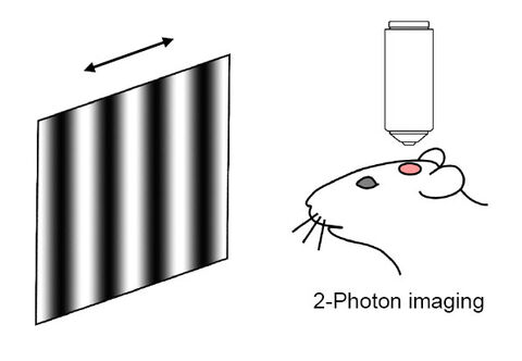
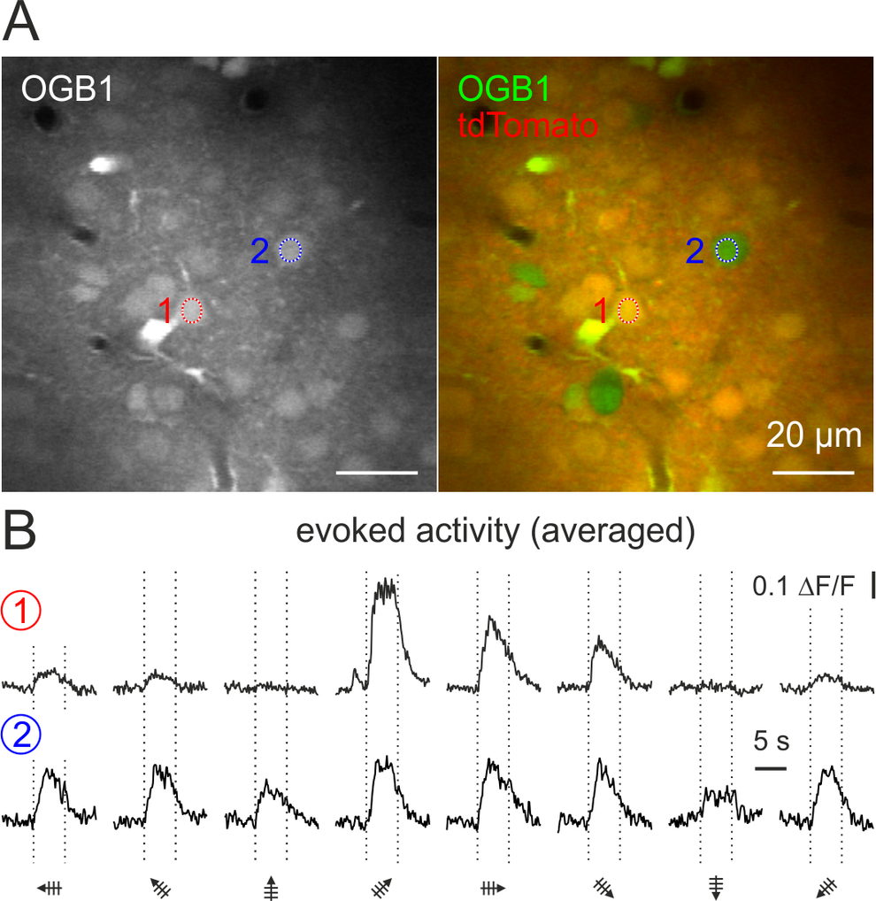
Wir verwenden die 2-Photonen Fluoreszenzmikroskopie in vivo, um die neuronale Netzwerkaktivität nach sensorischer Stimulation zu registrieren. Dabei dient die zellspezifische Expression von fluoreszierenden Proteinen der Identifikation verschiedener Zelltypen. So können während der Messung hemmende von erregenden Neuronen unterschieden werden (siehe Abbildung). Einzelne Zellen zeigen ein spezifisches Antwortverhalten auf bestimmte Eigenschaften des Stimulus, wie z.B. die Orientierung des präsentierten Streifenmusters.


Abb.: Auf der linken Seite ist der Versuchsaufbau schematisch dargestellt. Während der sensorischen Stimulation mit beweglichen Streifenmustern wird die neuronale Netzwerkaktivität im visuellen Kortex mit Hilfe der 2-Photonen Mikroskopie optisch abgeleitet. Auf der rechten Seite ist das Antwortverhalten eines erregenden Neurons (rote Eins) einem hemmenden Interneuron (blaue Zwei) gegenübergestellt (A). Während das erregende Neuron spezifisch nur auf bestimmte Orientierungen des präsentierten Streifenmusters antwortet, zeigt das hemmende Interneuron ein unspezifisches Antwortverhalten auf alle präsentierten Orientierungen (B).
Referenzen:
- Weiler S, Teichert M, Margrie TW (2024) Layer 6 corticocortical cells dominate the anatomical organization of intra and interhemispheric feedback. bioRxiv doi: https://doi.org/10.1101/2024.05.01.590702oRxiv
- Weiler S, Rahmati V, Isstas M, Wutke J, Stark AW, Franke C, Graf J, Geis C, Witte OW, Hübener M, Bolz J, Margrie TW, Holthoff K, Teichert M (2024) A primary sensory cortical interareal feedforward inhibitory circuit for tacto-visual integration. Nat Commun 15, 3081. https://doi.org/10.1038/s41467-024-47459-2
- Graf J, Samiee A, Flossmann T, Holthoff K and Kirmse K (2024) Chemogenetic silencing reveals presynaptic Gi/o protein-mediated inhibition of synchronized activity in the developing hippocampus in vivo. bioRxiv doi: https://doi.org/10.1101/2024.02.02.578550
- Graf J, Rahmati V, Majoros M, Witte OW, Geis C, Kiebel SJ, Holthoff K, Kirmse K (2022) Network instability dynamics drive a transient bursting period in the developing hippocampus in vivo. eLife 11:e82756. DOI: https://doi.org/10.7554/eLife.82756
- Weiler S, Rahmati V, Isstas M, Wutke J, Stark AW, Franke C, Geis C, Witte OW, Huebener M, Bolz J, Margrie TW, Holthoff K, Teichert M (2022) A primary sensory cortical interareal feedforward inhibitory circuit for tacto-visual integration. bioRxiv 2022.11.04.515161; doi: https://doi.org/10.1101/2022.11.04.515161
- Graf J, Rahmati V, Majoros M, Witte OW, Geis C, Kiebel SJ, Holthoff K, Kirmse K (2022) Network instability dynamics drive a transient bursting period in the developing hippocampus in vivo. bioRxiv doi: https://doi.org/10.1101/2021.05.28.446133
- Graf J, Zhang C, Marguet SL, Herrmann T, Flossmann T, Hinsch R, Rahmati V, Guenther M, Frahm C, Urbach A, Neves RM, Witte OW, Kiebel SJ, Isbrandt D, Hübner CA, Holthoff K, Kirmse K (2021) A limited role of NKCC1 in telencephalic glutamatergic neurons for developing hippocampal network dynamics and behavior. Proc Natl Acad Sci U S A. 2021 Apr 6;118(14):e2014784118. doi: 10.1073/pnas.2014784118
- Graf J, Zhang C, Marguet SL, Herrmann T, Flossmann T, Hinsch R, Rahmati V, Guenther M, Frahm C, Urbach A, Neves RM, Witte OW, Kiebel SJ, Isbrandt D, Hübner CA, Holthoff K, Kirmse K (2020) Intraneuronal chloride accumulation via NKCC1 is not essential for hippocampal network development in vivo. doi: https://doi.org/10.1101/2020.07.13.200014
- Kirmse K, Holthoff K (2020) Chloride transporter activities shape early brain circuit development, Book chapter in ´Neuronal Chloride Transporters in Health and Disease´, Academic Press, ISBN: 9780128153185
- Holthoff K (2020) The Janus-face of GABAergic synaptic transmission during brain development. J Physiol. doi: 10.1113/JP279623
- Zhang C, Yang S, Flossmann T, Gao S, Witte OW, Nagel G, Holthoff K, Kirmse K (2019) Optimized photo-stimulation of halorhodopsin for long-term neuronal inhibition. BMC Biology 17:95
- Kirmse K (2019) Editorial: GABAergic networks in the developing and mature brain. Brain Res. 1718:10-11. doi: 10.1016/j.brainres.2019.04.029
- Flossmann T, Kaas T, Rahmati V, Kiebel SJ, Witte OW, Holthoff K, Kirmse K (2019) Somatostatin Interneurons Promote Neuronal Synchrony in the Neonatal Hippocampus, Cell Rep 26(12):3173-3182.e5, https://doi.org/10.1016/j.celrep.2019.02.061
- Rahmati V, Kirmse K, Holthoff K, Kiebel SJ (2018) Ultra-Fast Accurate Reconstruction of Spiking Activity from Calcium Imaging Data. J Neurophysiol doi: 10.1152/jn.00934.2017.
- Kirmse K, Hübner CA, Isbrandt D, Witte OW, Holthoff K (2017) GABAergic Transmission during Brain Development: Multiple Effects at Multiple Stages. The Neuroscientist. DOI: 10.1177/1073858417701382
- Kirmse K und Holthoff K (2017) Funktionen der GABAergen Übertragung im unreifen Gehirn. Neuroforum 23(1):30–37.
- Kummer M, Kirmse K, Zhang C, Haueisen J, Witte OW, Holthoff K (2016) Column-like Ca2+ clusters in the mouse neonatal neocortex revealed by three-dimensional two-photon Ca2+ imaging in vivo. Neuroimage 138, 64-75. doi:10.1016/j.neuroimage.2016.05.050.
- Rahmati V, Kirmse K, Markovic D, Holthoff K, Kiebel SJ (2016) Inferring neuronal dynamics from calcium imaging data using biophysical models and Bayesian inference. PLoS Comput Biol 12(2): e1004736. doi:10.1371/journal.pcbi.1004736
- Kummer M, Kirmse K, Witte OW, Haueisen J, Holthoff K (2015) A method to quantify accuracy of position feedback signals of a three-dimensional two-photon laser-scanning microscope. Biomed Opt Express 6(10):3678-3693.
- Kirmse K, Kummer M, Kovalchuk Y, Witte OW, Garaschuk O, Holthoff K (2015) GABA depolarizes immature neurons and inhibits network activity in the neonatal neocortex in vivo. Nat Commun 6:7750, doi:10.1038/ncomms8750
- Hübner CA and Holthoff K (2013). Anion transport and GABA signaling. Front Cell Neurosci 7:177. doi: 10.3389/fncel.2013.00177
- Kummer M, Kirmse K, Witte OW, Holthoff K (2012) Reliable in vivo identification of both GABAergic and glutamatergic neurons using Emx1-Cre driven fluorescent reporter expression, Cell Calcium 52: 182-189.

