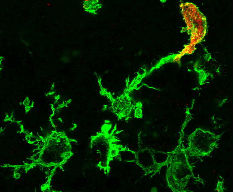
Macrophages are part of the innate immune system and essential for brain development and function. Using a novel method, scientists from Jena University Hospital, the University of Bonn and the Memorial Sloan Kettering Cancer Center in New York (USA) succeeded in visualizing macrophages that were formed in the bone marrow. In studies on mice, this technology enabled the researchers to observe that shortly after a stroke, numerous macrophages that had migrated from the blood begin to attack dead and adjacent healthy brain tissue. The results have now been published in the journal "Nature Neuroscience".
Jena/Bonn. In ancient Greek, the term macrophage means something like "big eater". These cells are components of our innate immune system that are present in every tissue of our body. In the brain, these resident immune cells are called microglia and play an essential role in normal brain development and function. During inflammatory processes, for example after stroke, brain infections, multiple sclerosis or Alzheimer's disease, other macrophages come into play, which are recruited from the bone marrow. Does it make a difference with regard to brain diseases whether the macrophages are resident in the brain or have migrated into the brain? This question has so far remained unresolved because scientists have not been able to differentiate between the two cell types using conventional methods.
This is precisely where the research of the working group led by Prof. Dr. Ralf Stumm from Jena University Hospital and his partners Prof. Dr. Elvira Mass from the University of Bonn and Prof. Dr. Frederic Geissmann from the Memorial Sloan Kettering Cancer Center in New York (USA) comes in. The team studied mice in which the genetic information for a molecular switch was introduced into a gene called Cxcr4. If this switch is activated with a special agent, stem cells in the bone marrow, from which all blood-circulating immune cells originate, produce a colored fluorescent protein.


